This article originally from here http://www.vet.uga.edu/vpp/clerk/gronfeld/index.php but it doesn’t exist any more.
Stacy Gronefeld, DVM; Holly A. Moore, DVM; Kenneth S. Latimer, DVM, PhD
Class of 2006 (Gronefield) and Department of Pathology (Moore, Latimer), College of Veterinary Medicine, University of Georgia, Athens, GA 30602-7388
Introduction
Canine rheumatoid arthritis is an erosive polyarthritis. It is a noninfectious, inflammatory, immune-mediated disease. Rheumatoid arthritis in dogs is not very common, and it has no sex predilection. It occurs mainly in small and toy breed dogs. Rheumatoid arthritis has been reported to occur in dogs from 8 months to 8 years of age, with the most common occurrence being 2 to 6 years of age. Rheumatoid arthritis is a chronic problem that can result in joint deformity.1,3,4
Etiology
The specific cause of rheumatoid arthritis is unknown. It has been speculated that canine distemper virus and the body’s immune response to this virus may play a role in the development of canine rheumatoid arthritis. Other possibilities are that type II collagen serves as an autoantigen4 and that some type of altered host immunoglobulin (IgG) is the inciting antigen that stimulates the immune response. Autoantibodies subsequently are formed and directed against the altered host immunoglobulin. These autoantibodies are called rheumatoid factors. The autoantibodies form complexes with the altered IgG molecules, and these immune complexes are deposited in the synovium of the joints. Inflammatory mediators are then activated, leading to a severe, erosive polyarthritis known as rheumatoid arthritis.3,4
Clinical Signs Associated with Rheumatoid Arthritis
Animals with rheumatoid arthritis often present with discomfort or pain in their joints. This can be seen as a shifting leg lameness or difficulty rising, walking up steps, and impaired ambulation. The joints that are most commonly affected are the carpal and tarsal joints. Affected joints may display signs of inflammation such as excessive warmth and/or swelling on palpation. Anorexia and malaise often are observed by the owner. The dog also may display a persistent fever. Splenomegaly and muscle wasting also have been reported with rheumatoid arthritis.1
Clinicopathologic Findings
Hemogram – A dog with rheumatoid arthritis can have a normal hemogram or may have leukocytosis, neutrophilia, and/or hyperfibrinogenemia. These changes reflect a generalized inflammatory process but do not lead to a specific diagnosis of rheumatoid arthritis.1
Synovial Fluid Analysis – Diagnostic indicators of rheumatoid arthritis usually are not present in the hemogram of diseased dogs. Synovial fluid analysis is more diagnostic of this condition. Arthrocentesis of multiple joints should be performed on a suspect animal. The complete synovial fluid analysis is composed of five major categories including physical appearance of the fluid, a mucin clot test, determination of protein concentration, performance of a nucleated cell count, and cytologic evaluation of the fluid.2 Serology also may be helpful if rheumatoid arthritis is suspected.
Physical Appearance
Normal synovial fluid should appear clear, colorless to pale yellow or straw colored, and lack turbidity. Viscosity also is evaluated with appearance. Synovial fluid is very viscous because it contains a high amount of hyaluronic acid. Normal viscosity is suggested when a strand of synovial fluid reaches 2 cm or greater before breaking. Viscosity also may be evaluated by examining a cytologic preparation of synovial fluid in which normal viscosity may cause cells to be arranged in rows. Rheumatoid arthritis is an inflammatory process and the number of cells in the synovial fluid will be increased. Increased cellularity causes the synovial fluid to appear more turbid than expected. Also, joint effusion dilutes the hyaluronic acid in the synovial fluid, causing a decrease in viscosity. Decreased viscosity is detected when the synovial fluid strand breaks before 2 cm in length. A suggestion of decreased viscosity also may be visualized on a smear when the cells are randomly distributed instead of appearing in rows.1,2
Mucin Clot Test
Mucin is hyaluronic acid. A mucin clot test is an assessment of the quality and quantity of hyaluronic acid. With this test, synovial fluid is expelled into 7N glacial acetic acid. The acetic acid causes the mucin to form a clot. Synovial fluid containing normal mucin will appear as clear fluid with a tight, ropy clot. Joint effusion from inflammation will dilute the hyaluronic acid, resulting in the formation of turbid fluid with a more friable mucin clot.2
Determination of Protein Concentration
Synovial fluid protein concentration is often measured by refractometry. Normal synovial fluid has a protein content of <1.0 g/dL (this value will be less than the bottom of the scale of most hand-held refractometers). Joint trauma and inflammation will increase the protein concentration of the synovial fluid. Therefore, an animal with rheumatoid arthritis will have an increased protein concentration in their synovial fluid.1,2
Nucleated Cell Count
Only nucleated cells are counted either by manual methods or automated analyzer. Healthy dogs may have a synovial fluid nucleated cell count of up to 1,500 cells/µl. Rheumatoid arthritis is an inflammatory process, causing an inflammatory synovitis. The total number of nucleated cells in the synovial fluid is moderately to markedly increased in an animal with rheumatoid arthritis.2
Cytologic Evaluation
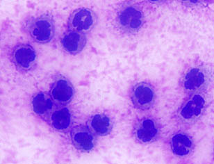
The most important part of the synovial fluid analysis is cytologic evaluation. Often, only a small amount of synovial fluid can be obtained. In this case, it should be used for cytologic study in preference to other forms of analysis. A wedge smear is made of the synovial fluid and stained with Romanowsky stain. Normal synovial fluid contains many mononuclear cells including macrophages and a few small, well differentiated lymphocytes (Fig. 1). Less than 10% of the total nucleated cell count consists of neutrophils.
Depending on the cellular content, synovial fluid can be classified as one of the following: normal, degenerative, inflammatory, or acute hemorrhage. Synovial fluid with inflammation can be further classified as either infectious or noninfectious, depending on the presence of microorganisms and the appearance of the neutrophils. Noninfectious inflammation in small animals is often associated with trauma or an immune-mediated process. In rheumatoid arthritis, the nucleated cell count is markedly increased (from >10,000 cells/µl to 100,000 cells/µl). The neutrophil is predominant cell type found in the synovial fluid (Fig. 2). Mononuclear cells also may be increased in number, but the neutrophil count alone can be >5,000 cells/µl.1,2

OLYMPUS DIGITAL CAMERA 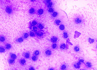

OLYMPUS DIGITAL CAMERA
Serology
In addition to routine synovial fluid analysis, serology also can aid in the diagnosis of canine rheumatoid arthritis. Serology will detect the rheumatoid factors (RF; autoantibodies directed against the altered IgG). A RF titer = 1:16 is considered a positive test result that is suggestive of rheumatoid arthritis. Positive serological tests are found in 20-70% of affected dogs; however, false positive test results can occur in dogs with other systemic inflammatory disorders. For this reason, a positive RF titer is not a definitive diagnostic test for rheumatoid arthritis. All of the clinical signs and diagnostic findings must be taken into account when interpreting the serological test.1,3
Summary
The earlier a diagnosis of rheumatoid arthritis can be made, the sooner treatment can begin. Early treatment is necessary to avoid irreversible damage to the joints. Medical treatment can involve the following: immunosuppressive drugs, gold salts, and/or nonsteroidal anti-inflammatory drugs. Aspirin has been used for palliative treatment. Immunosuppressive treatment usually involves the administration of prednisone and/or azathioprine or cyclophosphamide. Azathioprine is usually preferred as the second drug of choice because cyclophosphamide may have serious side-effects when used long-term. The dog should be re-examined and the synovial fluid should be re-evaluated 1 month after beginning treatment. Even with appropriate treatment, most dogs with rheumatoid arthritis will have a deterioration of their health status over time.3
Canine rheumatoid arthritis is an uncommon disorder in dogs. However, rheumatoid arthritis should be considered in any dog with a noninfectious, erosive polyarthritis. In addition to clinical signs, radiographic evidence of joint erosion, serologic testing, and laboratory analysis of synovial fluid may assist in disease diagnosis. Dogs with rheumatoid arthritis have synovial fluid that is thin and cloudy in appearance. Cytologically, rheumatoid arthritis is characterized by a noninfectious inflammatory appearance with the main cell type being neutrophils.
References
- Ettinger SJ, Feldman EC (eds): Textbook of Veterinary Internal Medicine, Diseases of the Dog and Cat, 5th ed. Philadelphia, W.B. Saunders Co., 2000, pp.78, 1879-1880, 1885.
- Latimer KS, Mahaffey EA, Prasse KW (eds): Duncan & Prasse’s Veterinary Laboratory Medicine: Clinical Pathology, 4th ed. Ames, Iowa State Press, 2003, pp. 318-321.
- Nelson RW, Couto CG (eds): Small Animal Internal Medicine, 2nd ed. St. Louis, Mosby, Inc., 1998, pp. 1070, 1085-1087, 1210.
- Tizard, IR: Veterinary Immunology, An Introduction, 7th ed. Philadelphia, W. B. Saunders Co., 2004, pp.405-408.